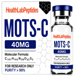

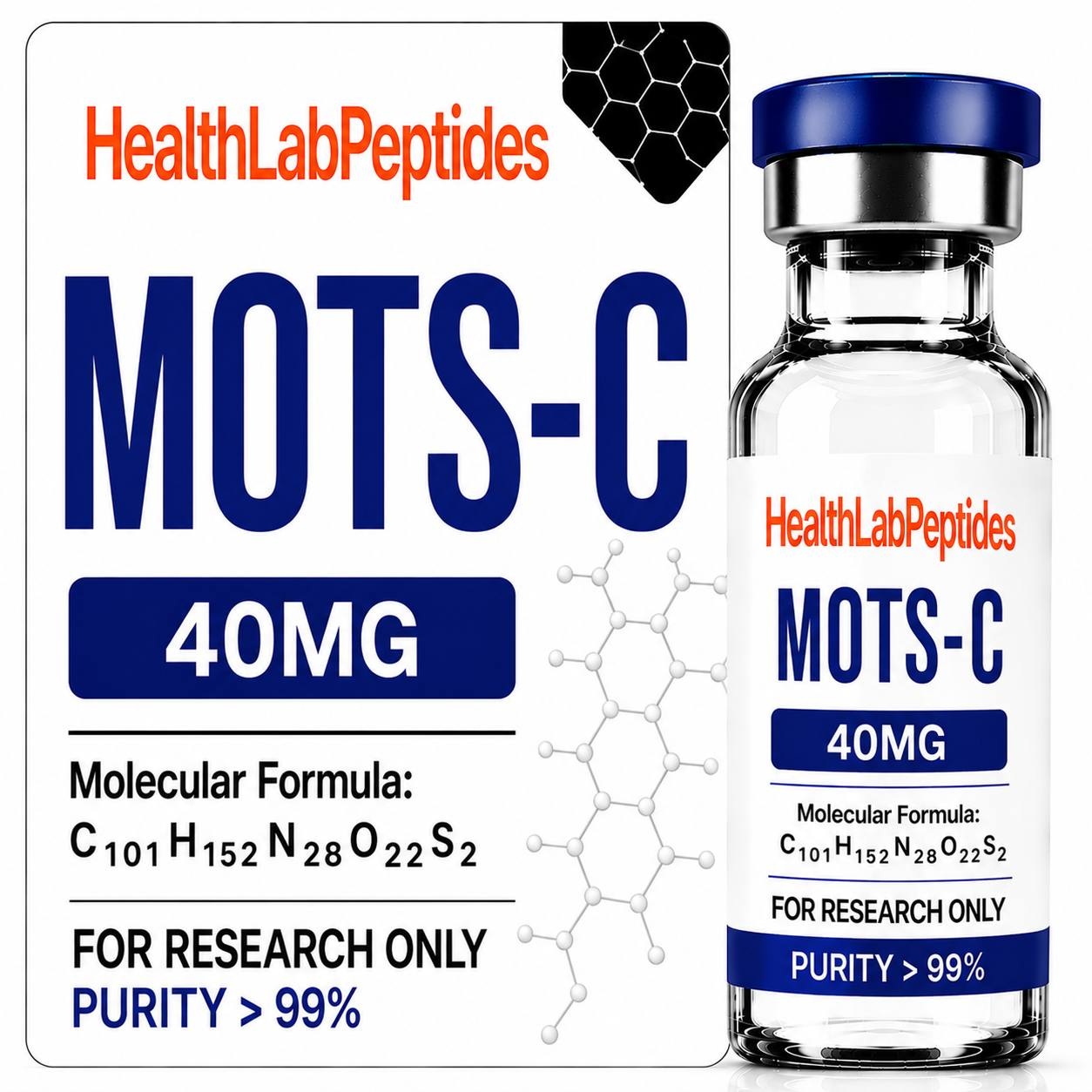
Research suggests that subcellular fractionation is an enrichment procedure, not automatic proof of mitochondrial localization. For U.S. laboratory teams, the practical question is not whether an instrument produces a number, but whether that number survives controls for identity, matrix, normalization, and analytical interference. Researchers reviewing MOTS-c 40 mg Research Peptide should treat the material as Research Use Only and define the measurement claim before collecting data.
Purity and recovery answer different questions
A fraction can be highly enriched yet recover only a small portion of the starting mitochondrial material. Conversely, high recovery may arrive with cytosolic, nuclear, endoplasmic-reticulum, or lysosomal contamination. Both purity and yield must be quantified relative to the starting homogenate and interpreted with a panel rather than a single marker.
For this laboratory question, the MOTS-c 40 mg Research Peptide product reference belongs directly in the experimental context: confirm lot identity, matrix recovery, and the exact in-vitro endpoint before comparing results.
Build the experiment around a falsifiable question
Choose markers representing the mitochondrial compartment of interest and the most plausible contaminating organelles. Analyze equal starting-material equivalents as well as equal protein loads, because these views answer different questions. A useful design separates the proposed biological event from assay mechanics. Include a vehicle control, a method blank where appropriate, a positive control with a known direction of response, and a negative control that challenges specificity. Predefine the primary endpoint and the rule for excluding failed wells or runs before viewing group labels.
Control the largest sources of false signal
Mechanical disruption, centrifugation force, temperature, delay before processing, organelle breakage, and pellet washing alter both recovery and cross-contamination. Record passage number, confluence, plate position, incubation interval, instrument settings, acquisition order, and operator. Randomization across plates and balanced processing order matter because drift can imitate a concentration trend. When fluorescence or luminescence is used, test the research material in a cell-free detection mixture to identify quenching, autofluorescence, or direct reporter effects.
Use orthogonal evidence
Combine immunoblot or targeted-proteomics marker panels with an independent functional or morphological check. Spike-recovery experiments can reveal whether the analyte is lost during isolation. Orthogonal does not simply mean repeating the same chemistry on another instrument. It means using a measurement with a materially different failure mode. Browse the HealthLab Peptides research catalog only after defining the assay, acceptance criteria, and documentation needed for the study.
A controlled comparison may include the SS-31 Research Peptide as a separate RUO research material, provided concentration, timing, analytical recovery, and assay conditions are matched rather than assumed equivalent.
- Verify research-material identity and lot traceability.
- Establish matrix recovery and linearity across the intended range.
- Include at least one specificity-challenging control.
- Normalize to a denominator justified by the biological question.
- Report independent experiments, not only technical replicate wells.
Interpretation boundaries
Detection in an enriched pellet supports association with that fraction under the stated protocol; it does not prove residence inside a particular mitochondrial membrane or matrix compartment. A statistically separated group is not automatically a mechanism, and an in-vitro response is not evidence of clinical utility. Report effect size, uncertainty, individual experiment values, and failed-quality-control events. Avoid translating cell or biochemical findings into human-use language.
Related RUO comparisons
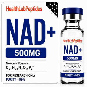
Depending on the hypothesis, researchers may compare the workflow with SS-31 Research Peptide or NAD+ Research Material. Such comparisons should be matched for analytical recovery and experimental context; nominal concentration alone does not establish equivalent exposure or activity.
A reproducible reporting checklist
Report rotor type, relative centrifugal force, duration, buffer composition, wash steps, starting equivalents, marker enrichment, and total recovery. Archive raw files, analysis scripts, plate maps, calibration results, reagent lots, and deviations. State exactly what the assay demonstrates and what it cannot resolve. This discipline makes a research-peptide article useful to laboratories without implying therapeutic, dosing, or consumer use.
Where the hypothesis requires a third product-specific comparator, the NAD+ Research Material should be evaluated with the same blanks, positive controls, interference checks, and independent replication.
Research Use Only disclaimer: This material is provided solely for lawful laboratory research and in-vitro analytical discussion. The compounds discussed are not medicines, foods, cosmetics, or consumer products and are not intended for human or veterinary use, ingestion, injection, diagnosis, treatment, cure, mitigation, or prevention of any disease. Nothing here is medical advice or a dosing, reconstitution, or administration guide. Qualified researchers are responsible for institutional review, applicable law, validated methods, appropriate controls, and safe laboratory handling.