

Introduction
Research suggests: glutathione participates in cellular redox biology through the chemical behavior of small glutathione molecules and through modifications of specific proteins. Protein S-glutathionylation, in particular, involves a reversible disulfide linkage between glutathione and a protein cysteine residue. A measurement of total reduced glutathione does not disclose which proteins were modified or what the consequences were. This article distinguishes GSH, GSSG, glutaredoxin, GSTO1, glutathione reductase, and the experimental readouts used in protein-thiol studies. The glutathione versus NAD+ research guide explains the related pyridine-nucleotide cofactors. This discussion is strictly for laboratory interpretation, without personal-use or health claims.
Why glutathione is chemically distinctive
Glutathione consists of the amino acids glutamate, cysteine, and glycine, arranged with an unusual gamma-glutamyl linkage. The cysteine thiol participates in important redox reactions and can form disulfide linkages. The reduced glutathione form is commonly called GSH, while the oxidized glutathione dimer is called GSSG. These forms represent different molecular species, and reporting them accurately is essential in scientific records. Measuring their amounts gives one perspective on an experiment, but does not reveal the modification state of a particular cellular protein. An inference about a named protein requires an assay that identifies that protein and its chemical state.
What protein S-glutathionylation means
S-glutathionylation involves the formation of a mixed disulfide between glutathione and a protein cysteine side chain. Depending on the protein and local biochemical conditions, this modification can influence enzyme activity, interactions, or conformation. It is a regulatory phenomenon rather than a universally harmful or beneficial event. A single cell may contain many proteins with different susceptible cysteine residues, and only some may respond to a particular experimental perturbation. An article that equates glutathionylation with general detoxification would conceal the main scientific question: which residue changed, how reliably was the change measured, and what functional consequence was demonstrated?
Specific residues versus bulk redox ratios
Bulk GSH/GSSG values summarize selected small-molecule measurements from a preparation; protein-site measurements ask whether glutathione was attached to a particular cysteine. These endpoints need not move together. A protein modification may change even when bulk glutathione measures change little, and a bulk redox shift does not identify its molecular targets. Differences among cytosolic, mitochondrial, and other cellular compartments further complicate interpretation. Scientists comparing papers should therefore record both the assay type and the sample compartment. Statements such as improved redox balance do not substitute for a description of the directly measured marker.
Glutaredoxin 1 as a distinct research enzyme
Glutaredoxin 1, usually abbreviated Grx1, is involved in certain protein-thiol redox reactions, including removal of glutathione modifications from protein substrates. The presence of glutaredoxin activity is not the same measurement as the amount of reduced glutathione in a cell. A laboratory report may test whether a particular substrate is modified and whether Grx1 can reverse that state. Demonstrating reversibility supports a molecular mechanism, but it does not establish that changing the system would be clinically beneficial. The experimental model, substrate identity, and control proteins determine how widely the result can be interpreted.
Glutathione reductase is a separate enzyme
Glutathione reductase participates in the reduction of GSSG to GSH using NADPH, not the same enzyme reaction studied when researchers test removal of protein S-glutathionylation by Grx1. The names are similar enough to cause confusion, but they refer to different enzymatic functions. Glutathione reductase measurements focus on a small-molecule redox cycle, while a protein-deglutathionylation study addresses a particular protein modification. This difference should be maintained in both figure captions and article prose. The peptide chemistry overview offers broader context on how short peptides differ from the larger enzymes that interact with them.
A 2023 biochemical review
Oppong, Schiff, Shivamadhu, and Ahn published “Chemistry and biology of enzymes in protein glutathionylation” in Current Opinion in Chemical Biology in 2023 (PMID 37245422, PMCID PMC10524987, DOI 10.1016/j.cbpa.2023.102326). The review discussed GSTO1, Grx1, and emerging LanC-like protein research. It also described how chemical inhibitors and biochemical probes helped establish substrate-specific mechanisms. A review organizes findings from multiple models, but it is not itself a clinical trial. Its discussion of enzymes associated with disease-relevant pathways does not demonstrate that a research-material glutathione product prevents or treats any condition in people.
Why GSTO1 cannot be replaced with a generic antioxidant label
Glutathione transferase omega 1, or GSTO1, has been investigated in relation to reactions involving specific protein substrates. The 2023 review discusses evidence for deglutathionylation of proteins such as NEK7 in some cellular systems. A named substrate provides a mechanistic question that is far more precise than saying an antioxidant system was activated. Even where a signaling effect is observed, attributing the entire biological response to GSTO1 requires appropriate controls and consideration of other pathways. The correct conclusion remains attached to the tested protein, cell model, and analytical evidence.
Why LanC-like proteins broaden the picture
Research on LanC-like proteins has raised questions about carbon-linked rather than sulfur-linked glutathione modifications in some settings. That distinction underscores the importance of chemical specificity: not every glutathione-associated attachment is the same bond. The term glutathionylation may encompass mechanistically different reactions in the literature, so study comparisons should inspect the original chemical characterization. Where identification is preliminary, it should be described as such. New biochemical categories can enrich research questions without supporting claims about any broad physiological improvement.
Methods used to detect modified proteins
Protein S-glutathionylation may be investigated using biochemical enrichment, antibody-based assays, mass spectrometry, or other analytical strategies. Each technique has limits. An antibody signal can suggest the presence of a modification without locating the exact residue. Mass spectrometry may help identify a candidate site but requires careful interpretation of fragments, confidence, and sample handling. Functional assays answer a different question: whether the identified modification changes protein behavior. A credible mechanistic explanation should distinguish identification, quantification, and functional validation instead of presenting one positive signal as proof of a complete pathway.
Sample handling and redox artifacts
Thiol-associated chemistry can change during collection or processing of a biological sample. This creates an important source of uncertainty: a detected molecular modification may not exactly represent the state that existed before the sample was handled. Scientific papers should explain how the investigators evaluated that risk and whether controls support the interpretation. Results from laboratories using different extraction and detection approaches may not be directly comparable. An evidence-based review describes this limitation rather than flattening every redox result into a single list of purported benefits.
Why time and cellular compartment matter
A redox modification may occur rapidly and then reverse, or may differ among cell populations and cellular compartments. A measurement at one time point captures only a snapshot. Cell lines and tissue preparations also differ in enzyme expression and access to substrates, which can produce distinct profiles. When comparing two reported findings, the investigator should ask which protein was measured, where the signal came from, and when material was collected. These details can explain differences without requiring a claim that one experiment disproved another. Mechanistic conclusions should match the resolution of the original observations.
Glutathione research compared with consumer terminology
Online descriptions frequently mention antioxidant support, immune health, detoxification, or protection. Those labels are not laboratory endpoints and should not be presented as established effects of RUO research material. The scientific findings discussed here concern named chemical species, protein modifications, and enzymatic reactions. Even if a biochemical mechanism relates to a disease pathway in a review, that relationship does not validate administration of a separate commercial reagent. Accurate SEO can address the underlying research question without using speculative health promises.
The HealthLab glutathione listing
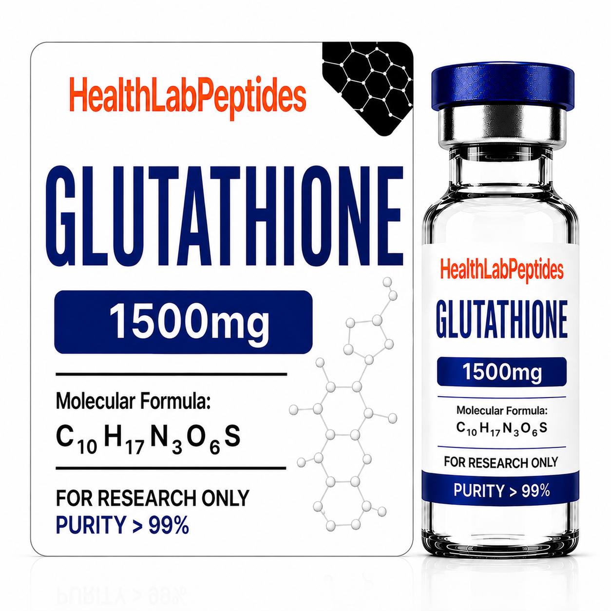
The HealthLab glutathione 1500 mg research material page identifies the catalog subject for lawful analytical work. A labeled quantity describes a product; it is not a protocol or a biological outcome. An available Certificate of Analysis may document selected batch findings, but cannot establish clinical safety, therapeutic efficacy, or suitability for human or veterinary use. Scientific literature about a molecule is a separate body of evidence from analytical documentation about a particular research-material batch. Researchers must preserve that distinction.
Frequently asked research questions
Is glutathione a peptide? Yes; it is a chemically distinctive tripeptide. Does GSH equal GSSG? No, they are different redox forms. Is glutaredoxin the same enzyme as glutathione reductase? No. Does S-glutathionylation always mean cellular injury? No, it can participate in regulation. Does a bulk glutathione ratio identify a modified protein? No. Can an isolated enzymatic finding prove a health outcome? No. Each question requires a model-specific measurement and carefully limited inference.
Research sources and conclusion
Primary reference for this discussion: Oppong and colleagues, Current Opinion in Chemical Biology (2023), PMID 37245422 and DOI 10.1016/j.cbpa.2023.102326. Other glutathione redox literature covers complementary biochemical reactions and measurement challenges. Glutathionylation research is most informative when the chemical bond, protein substrate, assay, and evidence level are stated explicitly. Protein-specific findings should not be converted into claims that an unapproved research-only material provides protection or treatment in people. The legitimate value of this article is its molecular interpretation.
Research Use Only
Research Use Only / Not for human or veterinary use. This article and any referenced HealthLab material are for lawful laboratory, analytical, and in-vitro research only. No dosing, preparation, administration, medical, cosmetic, or personal-use advice is given. These research materials are not represented as FDA-approved medicines.
 TB-500 Research Material — 5mg